Lumpy Skin Disease: An Emerging Disease In India And The Sub-Continent
Lumpy Skin Disease (LSD) was first identified in 1929 in Zambia after which it became endemic in many African countries. Of late the disease has been recorded and confirmed even in India.
Causes of Lumpy Skin Disease in Cattle
The disease is caused by a viral agent commonly called Lumpy Skin Disease Virus (LSDV). It belongs to the poxvirus family and bears similarities to sheep and goat pox. The virus is very resilient in that it can survive even in harsh temperature and is inactivated only above 55oC if exposed for 2 hours. It can be recovered from skin nodules kept on –80°C for 10 years and infected tissue culture fluid stored at 4°C for 6 months. It is also resilient to a wide pH range pH 6. 6–8. 6. It is susceptible to ether (20%), chloroform, formalin (1%). It is also susceptible to phenol (2%/15 minutes), sodium hypochlorite (2–3%), quaternary ammonium compounds (0.5%).
The virus remained for long periods in dried lesions, very resistant to inactivation, and can survive in necrotic skin nodules more than a month. The virus is inactivated in sunlight but can survive for many weeks if the barn is devoid of sunlight and dark. It is therefore difficult to get rid of the virus once there is an outbreak.
Morbidity rate varies between 10 and 50%, whereas mortality is low less than 5%. Although the virus belongs to the sheep/ goat pox family, this virus is very host-specific, can cause disease only in cattle and water buffalo. The morbidity and mortality, however, are higher in crossbreds compared to indigenous cattle. The virus has not been reported from wildlife. The virus is not known to be transmitted to humans.
Mode of Transmission
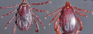
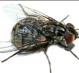 The main route of transmission is barthropod vectorsor, mosquitoes, biting flies, and male ticks. Transmission through the feed, water, and direct contact with infected skin is not known to be an important route of transmission. For transmission to occur the virus must be injected into the skin. Needle transmission from infected to clean animal there is a possibility. The virus is present in abundance in skin lesions, blood, saliva, ocular discharge for more than 35 days. The virus is also excreted in the semen of infected bulls, but the role of semen in the transmission of the disease is not known. There is only one report of transplacental transfer of the virus. It does not exhibit latency and the recrudescence of disease does not occur.
Diagnosis of lumpy skin disease
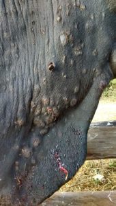
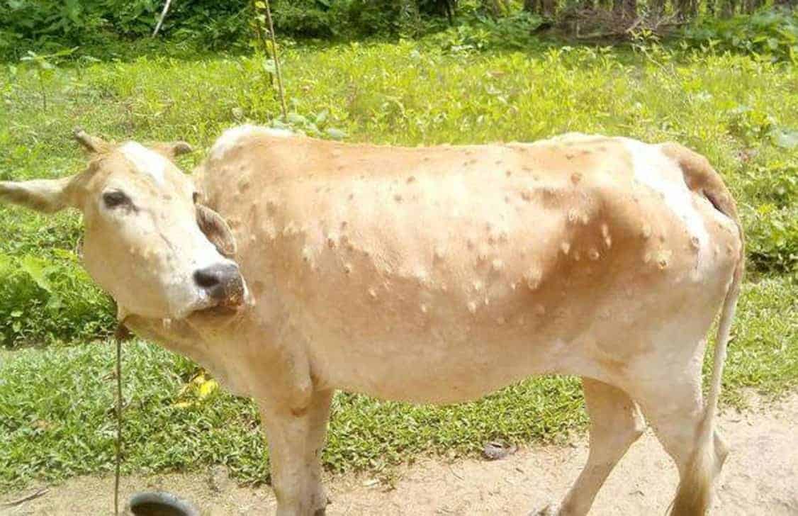
Diagnosis is not a big challenge as the symptoms are very typical. Initially, there is high fever that may exceed 41°C and in the case of lactating animals, there is a reduction in milk. The animal appears severely depressed, loss of appetite, and rapid loss of weight. In some cases rhinitis, conjunctivitis, excessive salivation, swollen lymph nodes. The typical lesions are cutaneous nodules of 2–5 cm in diameter particularly on the head, neck, limbs, udder, genitalia, and perineum within 48 hours of the onset of the febrile reaction. These nodules are firm to touch, round, and raised deep into the skin, subcutaneous tissue. The nodules become necrotic and can be observed for several months. Like pox lesions, the scars may remain for a long time, especially if the lesions were deep.
In a few cases, limbs and other ventral parts of the body, such as the dewlap, brisket, scrotum, and vulva, may be oedematous, causing difficulty in movement. If the bulls are infected these may become permanently or temporarily infertile. In the case of pregnant animals, abortion and temporary infertility may also be recorded. When the lesions are severe it may take a long time to recover due to emaciation, and secondary complications such as mastitis, pneumonia, and necrotic skin plugs. The disease may be confused with herpes mammillitis and cowpox, dermatophilosis, demodicosis, severe generalized insect bites.
Laboratory diagnosis: For confirmation of the diagnosis PCR is, ideal, for which skin nodules, scabs, saliva, nasal secretions, and blood are suitable samples.
Treatment and Control
It is a viral disease, hence there is no specific treatment. To take care of the secondary infection and severe inflammatory response non-steroidal anti-inflammatory drugs may be administered.
Early detection and rapid vaccination coverage are keys to effective prevention of the outbreak. As the virus is transmitted through insect bites, vector control is critical. In a small-hold system, ensuring that every time a sterilized needle is used only for a single animal. “Homologous” LSDV live attenuated vaccine strain, for example, “Neethling” LSD strain, “Heterologous” sheep pox or goat pox virus live attenuated vaccine strain.
Read: Organic Phosphinate Compounds Are Ineffective in Treating Phosphorous Deficiency in Animals